

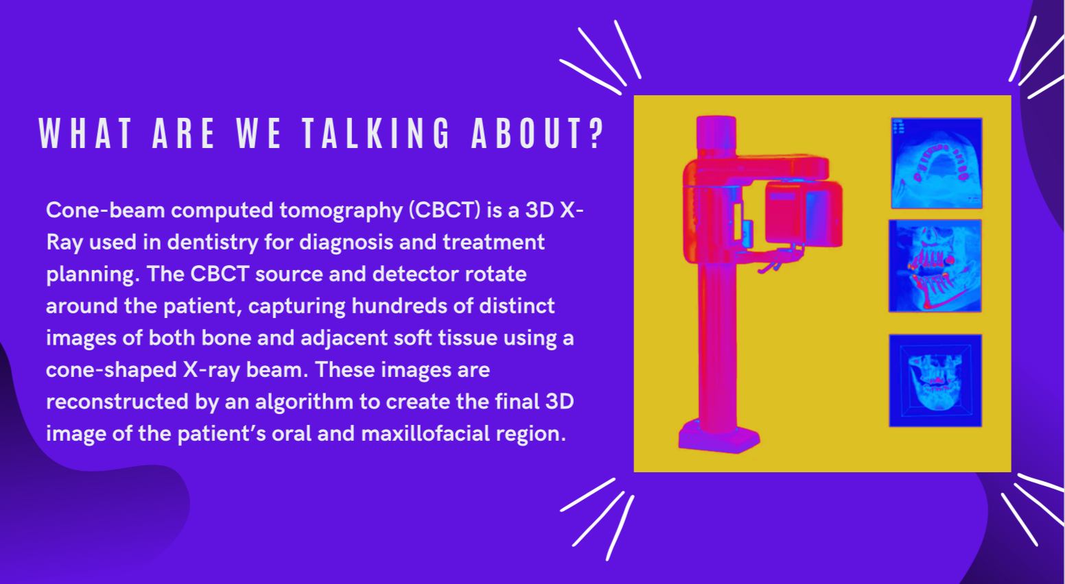
CBCT


Welcome back! On The Cusp is excited to explore Cone-Beam Computed Tomography (CBCT) technology. Read along with us as we share what we have learned about CBCT and how it can be a great tool in dental practice. Then, get ready for our guest speaker, Dr. Barzilay, who will discuss CBCT use in a Prosthodontics clinic!
For us techies, CBCT can also be combined with the workflow of other digital dentistry technologies, such as intraoral scanning, to enhance diagnosis and planning.


Did you know... CBCT differs from a traditional CT scanner.
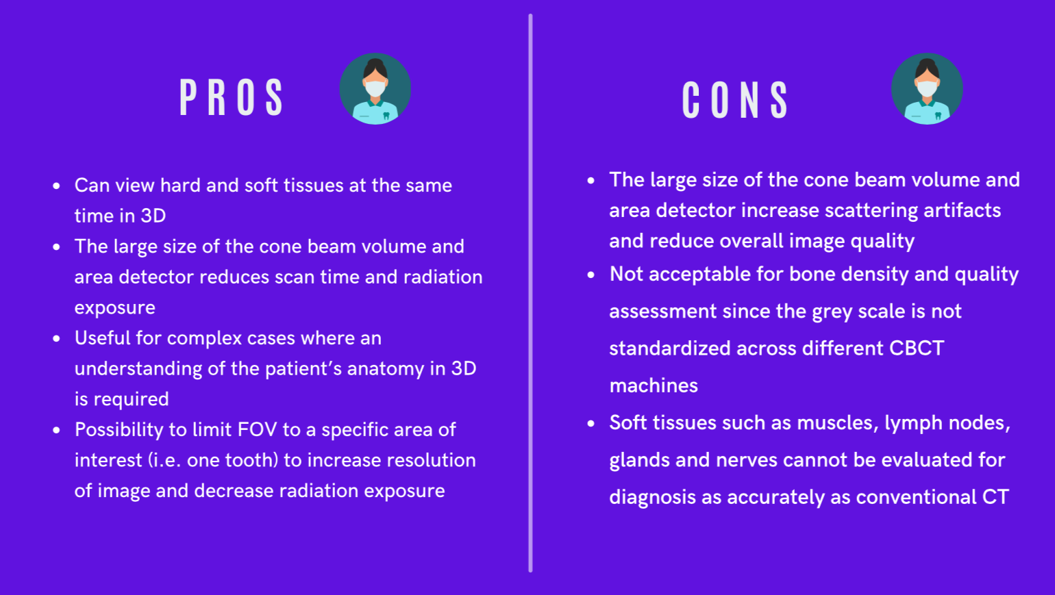
The “cone” in CBCT describes the shape of the X-Ray beam being delivered. This is different from the fan shape beam delivered by conventional CT scanners! Also, an important difference is that the radiation exposure from CBCT is up to 10 times less than that incurred from conventional CT scanning. However, the soft tissues, such as muscle or lymph nodes images generated by CBCT are not as comprehensive for diagnostic purposes as conventional CT.
To better visualize this technology, check out this short video!


P.S. future-self:
Please note, this imaging technique delivers higher levels of radiation than traditional 2D X-Rays, so let’s remember our ‘ALARA’ principle (As Low As Reasonably Achievable). In other words, CBCT should be used only when necessary to provide clinical information that can’t be obtained using other imaging technologies.

E. Voorand ~ S. Roberge ~ R. Brunswick ~ DDS 2024s
Sources Consulted
https://www.ncbi.nlm.nih.gov/pmc/articles/PMC4377156/
https://perdidobaydental.com/difference-dental-x-ray-ct-scan
https://www.ncbi.nlm.nih.gov/pmc/articles/PMC3520262/
https://en.wikipedia.org/wiki/Cone_beam_computed_tomography
https://agd.org/constituent/news/2020/02/17/cbct-purchasing-guide-how-to-choose-the-perfect-machine
https://www.nibib.nih.gov/science-education/science-topics/computed-tomography-ct
https://www.radiologyinfo.org/en/info/dentalconect https://www.ncbi.nlm.nih.gov/pmc/articles/PMC3520262/
http://cosmodont.com/dental-services/cbct-3d-scan